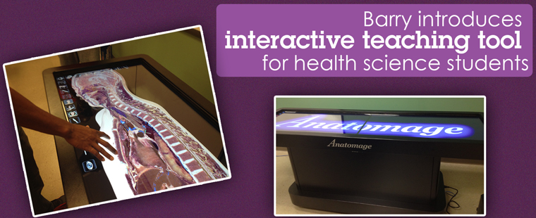
Barry University has recently acquired new technology that allows current and future physicians to examine the human body like never before through the use of life-sized, virtual anatomy imagery. Like a gigantic tablet, the Anatomage table allows students and faculty to virtually dissect the human anatomy layer by layer. They can customize various pathologies and visualize skeletal tissues, muscles, organs, and soft tissue. Barry is one of only four schools in Florida, and the only school in Miami-Dade County, to own the new equipment.
“We are excited to be able to offer this state-of-the-art learning tool to our students,” said Dr. John McFadden, CRNA dean of the College of Nursing and Health Sciences at Barry. “Very few universities have this technology in the classroom. It certainly gives Barry’s Biomedical Science students a competitive edge in how they learn anatomy and pathology.”
This new technology allows for medical schools to have an alternative to traditional cadavers. Supporters of the 3-D virtual technology say inexperienced students can practice their skills and redo their incisions in a way that real cadavers do not allow. This allows medical students to further sharpen their skills by the time they are ready to work on a real person.
The same holds true for experienced physicians, who can use scans of actual patients and inspect body images before surgery to minimize any surprises during an operation. They can see what is there before making the first incision. Surgeons can also use the Anatomage table to locate specific pathology in several views and decide treatment plans and post-surgical reviews.
“While I believe that real cadavers will always be the standard tool teaching anatomy to medical students, the Anatomage table provides an excellent supplement,” said Dr. Sathees Chandra, director and professor of the Biomedical Sciences program at Barry. “It allows us to teach in a new and creative way.”
Unlike plastic models, the table allows students to view structures at infinite angles and better understand how different structures relate within the body. The table offers realistic visualization of 3-D anatomy by combining imaging from X-rays, ultrasounds, and MRIs.
The Anatomage table costs approximately $80,000 and can hold up to a terabyte of data, which equals information for about 1,000 patients.